BET sorptometer for N2 Ar and Kr BET surface areas and A microporosities organic carbon analyzer. The lab is critical to a.
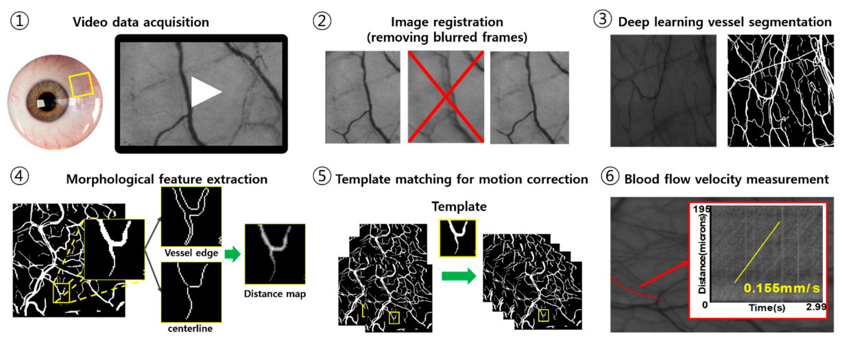
Sensors Free Full Text Quantification Of Blood Flow Velocity In The Human Conjunctival Microvessels Using Deep Learning Based Stabilization Algorithm Html
High Energy Theory including Cosmology.
. Patient-specific case studies to quantify relative bounds to this uncertainty uncovered a dire need for care in the virtual model reconstruction from medical images. Additionally we have a powerful suite of lasers and cameras for advanced laser-based imaging. The Structural Diagenesis Initiatives main microstructural imaging instruments are housed in the Bureau of Economic Geologys Core Research Center in north Austin.
Scanning Electron Microscope SEM Lab. AI Health Lab is made up of scholars and students from different fields and disciplines. Provides micro to nanoscale SEM imaging chemical analysis of various rock types.
The goal of the airborne imaing effort at UT Austin is to develop applications of new remote sensing technology to map coastal near shore environments and landscape or lacustrine features outside the coastal zone. Learn More About the Department. Together these resources provide opportunities to utilize a variety of approaches including cell culture.
Our lab facilities include space for parasite rearing and experimental infections microscopes for dissections to enumerate parasite or measure morphology and the essentials for genetics genomics and some immunology. The facilities of the Flowfield Imaging Lab are located at the JJ. We study the state and.
Center for Global Innovation and. UT GeoFluids is managed by the University of Texas Institute for Geophysics UTIG and is currently supported by 10 energy companies at a cost of 50000year. UT Austin offers hundreds of acres of biological research stations.
Our work emphasizes the application and development of laser diagnostics to help us understand the physics of complex turbulent. Justin Rousseau from Dell Medical School at the University of Texas at Austin. Andrew Dunn 107 W.
Wednesday March 6 2019. Meanwhile the Center supports millions of specimens in its Biodiversity Collections housed at the Lake Austin Center JJ. Ying Ding from School of information and Prof.
The SEM lab contains our Zeiss Sigma field emission instrument and the FEI Nova Nano 430 Field Emission SEM. Lab space is adjacent to 9 other neuroscience research labs with shared facilities that include a Nikon Imaging Center flow cytometry and cell sorting core facility and a newly constructed vivarium in the building. We are at the start of a 10-year effort entitled GeoFluids2020.
Synthesis and strong magnetic field equipment. Biomedical Engineering Building 107 W. Atomic Molecular and Optical Physics.
Welcome to the Flowfield Imaging Lab. Welcome to Biomedical Engineering. Imaging LSCI is a well-established technique for full-field optical imaging of cortical blood flow.
Pickle Research Campus and on UTs main campus. The Microscopy and Imaging Facility also manages the Flow Cytometry Laboratory which houses several instruments capable of fluorescence-based cell analysis and cell sorting. In certain cases relative errors in the computed flow field due to geometric uncertainty can be of.
Our results are used to predict pressure and stress design stable and safe drilling programs and predict hydrocarbon migration and entrapment. Dynamic Light is based in Austin Texas and was founded in May 2018. The Laboratory Supplies office located in PMA 8306 471-5352 provides supplies for laboratory classes as well as providing more general office supplies for faculty and administrators within the Department.
In separate laboratory space the Core Research Center also houses the initiatives Fluid Inclusion Laboratory and. We are part of the Department of Aerospace Engineering and Engineering Mechanics at the University of Texas at Austin. These include cell sorting and analytical flow cytometry to phenotype quantify and isolate cells and confocal microscopy to capture high resolution imagesp Pamela Whitney - Lab.
We are looking to hire a talented young investigator for a post-doctoral fellowship in computational oncology within the U Follow utbiomedical. The Bolnick lab at UT Austin has two studentpostdoc offices a computer lab two aquarium rooms for our stickleback fish colonies and four lab rooms. Austin TX 78712 Mailing Address The University of Texas at Austin Biomedical Engineering Attn.
A national network of women deans chairs and distinguished faculty in biomedical engineering including from the Cockrell School of. The University of Texas High-Resolution X-ray Computed Tomography Facility. We do research and teaching in the primary areas of turbulent mixingcombustion and high speed shear flows.
Nuclear magnetic and electron paramagnetic. Dynamic Lights mission is to enable real-time blood flow imaging to improve patient care and lower health care costs. All shared core facilities elsewhere at UT Austin are available for use.
Biophysics Nonlinear Dynamics. Please contact a member of our staff for inquiries about instrument specifications or to schedule training. We focus on cutting-edge research on AI in health and data-driven science of science.
1 4 In vivo measurements of molecular oxygen have historically been made using highly invasive Clarke electrodes that are limited to point measure-ments outside the vascular lumen. The Computational Imaging Lab at UC Berkeley develops methods for designing imaging systems and algorithms jointly in terms of hardware and software. High Energy Particle Experiment.
Biomedical Imaging Center The University of Texas at Austin 100 East 24th St NHB Suite 0240 Austin TX 78712. This new generation of cameras integrates computers as a part of the imaging system whereby the optical setup and postprocessing algorithms are designed simultaneously. MDACC Science Park Molecular Biology Core.
AI Health Lab is led by Prof. The Center for Biomedical Research Support Microscopy and Imaging Center is dedicated to supporting the research community at UT Austin and beyond by providing access to state-of-the-art imaging instrumentation as well as technical assistance and education to researchers across all disciplines. Dean Keeton St Stop C0800 Austin TX 78712.
Noise and flow artifacts in the medical images and clinical measurements lead to geometric models that contain some uncertainty. For example one can digitally refocus images enhance resolution or. 5 7 Magnetic resonance tech-niques allow for noninvasive imaging of hemoglobin saturation but suffer from low.
Carbon analyzer TC Organic analysis Field and laboratory gas chromatographs thermal desorber high pressure liquid chromatographs Inorganic analyses Ion chromatograph autotitrator field and lab spectrophotometers. The High-Resolution X-ray Computed Tomography Facility at The University of Texas at Austin UTCT is a national shared multi-user facility supported by the Instrumentation and Facilities Program of NSFs Earth Sciences EAR directorateUTCT offers scientific researchers across the earth biological and engineering. At the Brackenridge Field Lab and Stengl Lost Pines students and researchers study the interactions between life and the land.
The Flow Cytometry and Cell Imaging Core Facility at Science Park provides cutting edge cell analysis resources. The main facilities include the High-Speed Wind Tunnel Mach 3 Wind Tunnel Turbulent Jet Flame Facility Combustion Wind Tunnel and the 125 Second Droptower. Modern facilities for graduate study and research include.
Our technology supports better decision making by providing surgeons continuous real-time and high-resolution visualization of blood flow and perfusion.

Amnis Imagestream X Mk Ii Imaging Flow Cytometer

Flowfield Imaging Lab Facilities Equipment

Imaging Flow Cytometry Reveals That Granulocyte Colony Stimulating Factor Treatment Causes Loss Of Erythroblastic Islands In The Mouse Bone Marrow Experimental Hematology

Amnis Imagestream X Mk Ii Imaging Flow Cytometer

Amnis Imagestream X Mk Ii Imaging Flow Cytometer

Amnis Imagestream X Mk Ii Imaging Flow Cytometer

Csf Flow Imaging In Chiari 1 Malformation Ppt Video Online Download

Fetal Ductus Arteriosus Constriction Related To Fluoxetine At Diagnosis Download Scientific Diagram
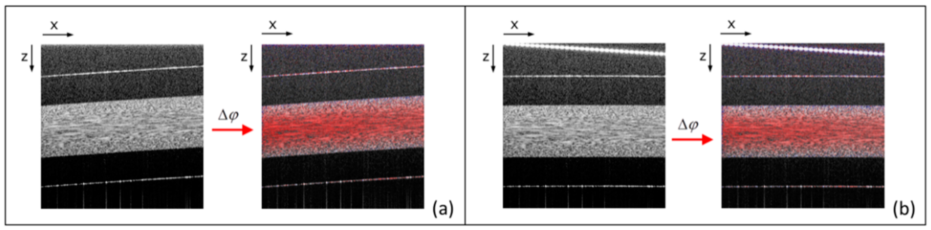
Applied Sciences Free Full Text Flow Measurement By Lateral Resonant Doppler Optical Coherence Tomography In The Spectral Domain Html
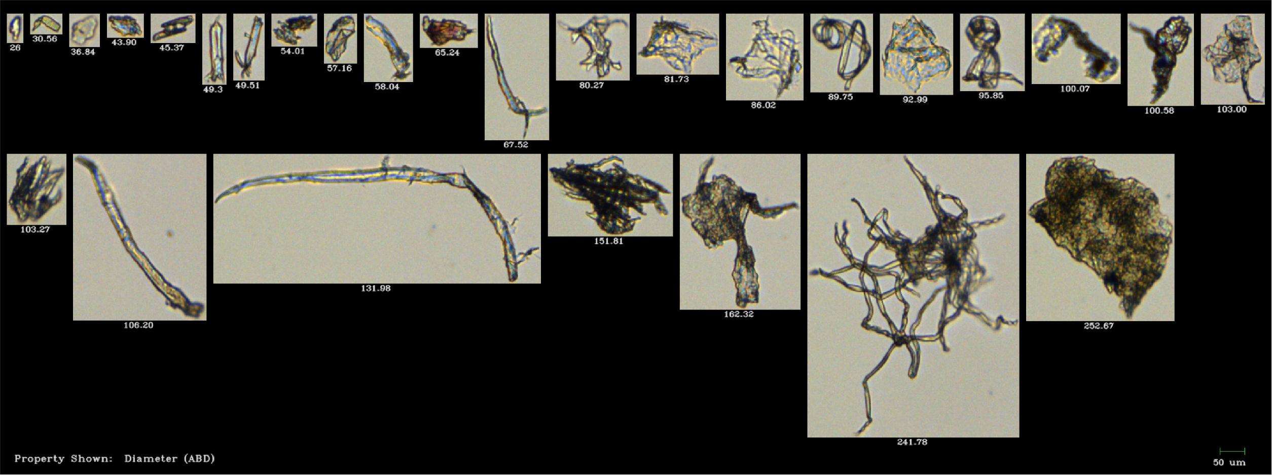
Microplastics In Our Oceans How Can We Study These Microscopic Pollutants

Microplastics In Our Oceans How Can We Study These Microscopic Pollutants

Amnis Imagestream X Mk Ii Imaging Flow Cytometer
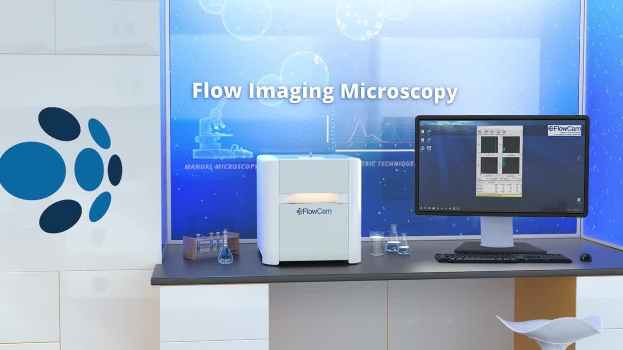
What Is Flow Imaging Microscopy
Applied Sciences Free Full Text Flow Measurement By Lateral Resonant Doppler Optical Coherence Tomography In The Spectral Domain Html

I Ve Made A Flowchart To Help Me Tell Amino Acids Apart Until I Know Them A Bit Better That Is Imgur Flow Chart Biochemistry Notes Biochemistry

What Is Flow Imaging Microscopy

Flowfield Imaging Lab Facilities Equipment

Flowfield Imaging Lab Members

Amnis Imagestream X Mk Ii Imaging Flow Cytometer


0 komentar:
Posting Komentar